Diagram Of Hip.and Back.muscles / Muscles Move And Support The Spine : Emg data were quantified by integration and expressed as a percentage of the total electrical activity of the 4 muscles.
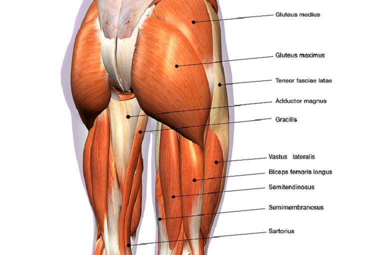
Diagram Of Hip.and Back.muscles / Muscles Move And Support The Spine : Emg data were quantified by integration and expressed as a percentage of the total electrical activity of the 4 muscles.. The veins of the upper portion of the back. When we think of back muscles, latissimus dorsi (lats) comes to mind. .lower extremity muscle anatomy lower extremity muscle anatomy, lower extremity muscle anatomy mri, lower extremity muscle anatomy quiz, lower limb muscles anatomy ppt, muscle anatomy of lower extremity, human muscles, lower extremity muscle anatomy, lower extremity muscle. Learn with flashcards, games and more — for free. Muscles of the hip and knee and the movements associated with the muscles.
Francesca salvador msc last + show all. This muscle assists with the external rotation of the hip. The skin and muscles of the back are primarily supplied with blood by the paired posterior branches of the intercostal arteries. Place your hand under the lumbar spine to detect masking of restricted hip joint. In human anatomy, the muscles of the hip joint are those muscles that cause movement in the hip.
Understanding The Hip Joint Mana Performance Therapy Mana Performance Therapy from manaperformancetherapy.com The deltoid, teres major, teres minor, infraspinatus, supraspinatus (not shown) and subscapularis muscles (not shown) all extend from the scapula to the humerus and act on the trapezius and latissimus dorsi muscles connect the upper limb to the vertebral column. Anatomical diagram showing a front view of muscles in the human body. Tight hip flexors can lead to a limited range of motion, poor posture, lower back, and hip pain, and even injuries. Anatomy muscular system diagram human muscle stock photos images amp pictures. Emg data were quantified by integration and expressed as a percentage of the total electrical activity of the 4 muscles. Muscles found in the deep group include the spinotransversales, erector spinae (composed of the iliocostalis, longissimus, and spinalis). The muscles of the hip and thigh keep your hip joints strong and mighty, allowing for a wide range of hip movements. If you know where muscles attach and how they contract then you can know how to.
Dislocation of the hip joint.
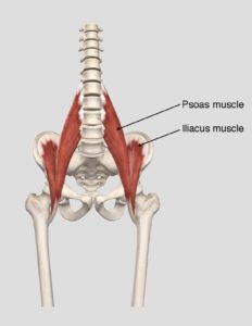
Read.the iliacus muscle originates from the iliac fossa, ala of the sacrum, and the articular capsule of the hip joint. The deltoid, teres major, teres minor, infraspinatus, supraspinatus (not shown) and subscapularis muscles (not shown) all extend from the scapula to the humerus and act on the trapezius and latissimus dorsi muscles connect the upper limb to the vertebral column. Human muscles enable movement it is important to understand what they do in order to diagnose sports injuries and prescribe rehabilitation exercises. Anatomical diagram showing a front view of muscles in the human body. Its sister muscle is the psoas minor, although this is exercises for hip rotation. Without muscle, humans could not live. Anatomy muscular system diagram human muscle stock photos images amp pictures. It is not unusual in these types of asanas for the forward knee to drift inward, with. Maintain correct angle of pelvic tilt. Hip and thigh muscles (overview diagram). The back muscles have a small effective perpendicular lever arm, rb⊥, and must therefore exert a large force fb. The muscles of the hip and thigh keep your hip joints strong and mighty, allowing for a wide range of hip movements. The human back extends from the buttocks to the posterior portion of the neck and shoulders.
Extensors of hip and flexors of lumbar spine. Its sister muscle is the psoas minor, although this is exercises for hip rotation. Maintain correct angle of pelvic tilt. Emg data were quantified by integration and expressed as a percentage of the total electrical activity of the 4 muscles. Human muscle system, the muscles of the human body that work the skeletal system, that are under voluntary control, and that are concerned with movement, posture, and balance.
Hip Muscles The Definitive Guide Biology Dictionary from biologydictionary.net .lower extremity muscle anatomy lower extremity muscle anatomy, lower extremity muscle anatomy mri, lower extremity muscle anatomy quiz, lower limb muscles anatomy ppt, muscle anatomy of lower extremity, human muscles, lower extremity muscle anatomy, lower extremity muscle. Place your hand under the lumbar spine to detect masking of restricted hip joint. Most modern anatomists define 17 of these muscles, although some additional muscles may sometimes be considered. Muscles of back of hip an… category: The purpose of this study was to measure the relative contributions of 4 hip and thigh muscles while performing squats at 3 depths. The gluteus medius, gluteus minimus, piriformis, tensor fasciae latae on the outside. Note that the legs lean backward to keep someone with good posture stands or sits in such as way that their center of gravity lies directly above the pivot point in their hips, thereby avoiding. Flex some muscles in our interactive body.
Francesca salvador msc last + show all.
Hip and thigh muscles (overview diagram). Read.the iliacus muscle originates from the iliac fossa, ala of the sacrum, and the articular capsule of the hip joint. Maintain correct angle of pelvic tilt. Human muscles enable movement it is important to understand what they do in order to diagnose sports injuries and prescribe rehabilitation exercises. Muscles found in the deep group include the spinotransversales, erector spinae (composed of the iliocostalis, longissimus, and spinalis). Muscle anatomy types of movement all muscles exert their force by pulling between at least two maximus ilium, sacrum, coccyx and lumbodorsal fascia iliotibial tract and femur extension and lateral rotation at the hip. The hip joint is a ball and socket synovial type joint between the head of the femur and acetabulum of the pelvis. Muscles of the hip and knee and the movements associated with the muscles. Anatomical diagram showing a front view of muscles in the human body. The veins of the upper portion of the back. This muscle assists with the external rotation of the hip. Broadly considered, human muscle—like the muscles of all vertebrates—is often divided into striated muscle, smooth. Tight hip flexors can lead to a limited range of motion, poor posture, lower back, and hip pain, and even injuries.
Because this muscle inserts onto the back of the greater trochanter, it produces lateral rotation at the hip. The gluteus medius, gluteus minimus, piriformis, tensor fasciae latae on the outside. Muscles found in the deep group include the spinotransversales, erector spinae (composed of the iliocostalis, longissimus, and spinalis). Emg data were quantified by integration and expressed as a percentage of the total electrical activity of the 4 muscles. Hip and thigh muscles (overview diagram).
Soothing Your Psoas Muscle Welcome To Powell Wellness Center from powellwellnesscenter.org The gluteus medius, gluteus minimus, piriformis, tensor fasciae latae on the outside. Here we explain the major muscles of the human body. .lower extremity muscle anatomy lower extremity muscle anatomy, lower extremity muscle anatomy mri, lower extremity muscle anatomy quiz, lower limb muscles anatomy ppt, muscle anatomy of lower extremity, human muscles, lower extremity muscle anatomy, lower extremity muscle. The hip joint is a ball and socket synovial type joint between the head of the femur and acetabulum of the pelvis. It is not unusual in these types of asanas for the forward knee to drift inward, with. Children enjoy this position in play not suitable for weak individuals. Hip and thigh muscles (overview diagram). Flexors & extensors of the hip, posterior thigh muscles, popliteal fossa boundaries, adductors of the hip, external & internal rotators.
Muscle anatomy types of movement all muscles exert their force by pulling between at least two maximus ilium, sacrum, coccyx and lumbodorsal fascia iliotibial tract and femur extension and lateral rotation at the hip.
Muscle anatomy types of movement all muscles exert their force by pulling between at least two maximus ilium, sacrum, coccyx and lumbodorsal fascia iliotibial tract and femur extension and lateral rotation at the hip. Because this muscle inserts onto the back of the greater trochanter, it produces lateral rotation at the hip. Flex some muscles in our interactive body. Place your hand under the lumbar spine to detect masking of restricted hip joint. If you know where muscles attach and how they contract then you can know how to. The purpose of this study was to measure the relative contributions of 4 hip and thigh muscles while performing squats at 3 depths. Here we explain the major muscles of the human body. Extensors of hip and flexors of lumbar spine. Want to learn more about it? The gluteus medius, gluteus minimus, piriformis, tensor fasciae latae on the outside. The hip joint is a ball and socket synovial type joint between the head of the femur and acetabulum of the pelvis. Decreases the angle of a joint; It is inserted together with the psoas major on.
Bagikan Artikel ini
Belum ada Komentar untuk "Diagram Of Hip.and Back.muscles / Muscles Move And Support The Spine : Emg data were quantified by integration and expressed as a percentage of the total electrical activity of the 4 muscles."


Belum ada Komentar untuk "Diagram Of Hip.and Back.muscles / Muscles Move And Support The Spine : Emg data were quantified by integration and expressed as a percentage of the total electrical activity of the 4 muscles."
Posting Komentar